
Understanding Your ECG: What That Squiggly Line Actually Means
*Collaborative Post
If you’ve ever had an ECG in a GP’s Clinic or the back of an ambulance, you’ve likely watched the machine spit out a strip of pink grid paper covered in jagged peaks and troughs and wondered what the clinician is looking for.
An electrocardiogram (ECG) is one of the most routine investigations in medicine. It takes roughly two to three minutes, costs nothing in discomfort beyond peeling off sticky tabs/stickers, and gives doctors immediate information about how your heart is behaving.
At the same time, it’s often misunderstood. A normal ECG doesn’t hand you an unconditional clean bill of cardiac health, nor does an ‘abnormal’ finding automatically mean you’re on the verge of a cardiac arrest.
Here is what the test actually measures, what it catches, and where its blind spots lie.
How It Works?
Every time your heart beats, an electrical current spreads from top to bottom, telling the muscular chambers precisely when to contract and pump blood. An ECG simply listens to that current.
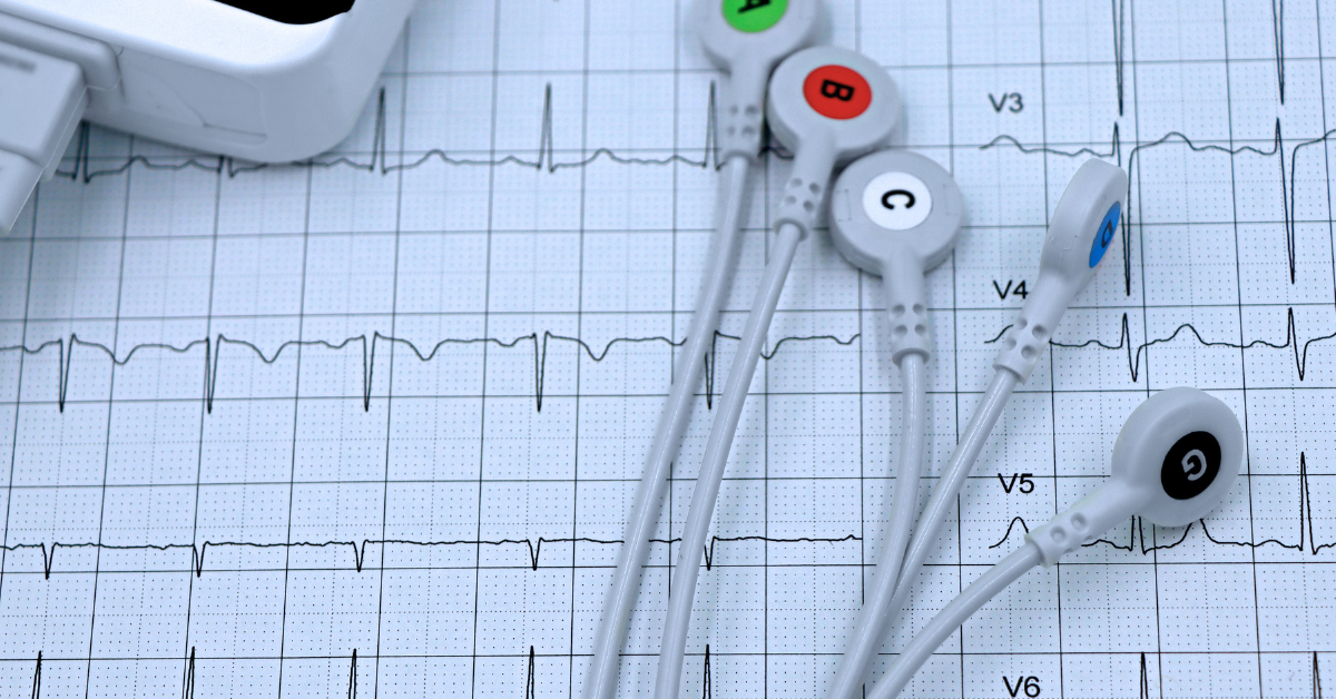
Small adhesive patches (electrodes) are placed on your chest, wrists, and ankles, usually 10 sensors in total. These record the electrical voltage your heart produces from 12 distinct angles (which is why clinicians call it a 12-lead ECG, referring to the viewpoints, not the number of wires).
It is a purely passive recording. The machine doesn’t emit any electricity, uses no radiation, and injects nothing into your body. Aside from the mild sting of the adhesive pulling on body hair when the pads come off, the test is entirely painless.
What a Doctor Looks for on the Strip?
The classic heartbeat on an ECG consists of a small bump (the P wave), a sharp spike (the QRS complex), and a final rounded wave (the T wave). Each represents a specific phase of the heart charging and discharging.
By measuring the height, width, and timing of these waves, your doctor answers core questions:
1. Is the rhythm regular or chaotic?
The machine shows whether beats are originating where they should (the sinus node) at a steady interval.
- Normal Sinus Rhythm: The standard, healthy electrical pattern.
- Atrial Fibrillation (AF): The upper chambers quiver erratically rather than contracting smoothly. AF is common, especially as we age, and matters because blood can pool and form clots, increasing the risk of stroke.
- Ectopic beats: Harmless ‘extra’ beats that feel like a flutter or a skipped beat in your chest. Almost everyone gets them; an ECG helps confirm they’re benign.
2. Is the rate appropriate?
A resting heart rate between 60 and 100 beats per minute (bpm) is standard. Below 60 is bradycardia (common and healthy in athletes, but sometimes a sign of conduction trouble in older adults). Above 100 is tachycardia, which can reflect anxiety, fever, dehydration, thyroid issues, or a primary rhythm disorder.
3. Is there evidence of an active or past heart attack?
When heart muscle is deprived of oxygen, its electrical properties change instantly. Certain patterns, such as an elevated ST segment, tell an phys that a coronary artery is blocked and needs opening immediately. The trace can also reveal permanent scars from previous, sometimes unnoticed heart attacks.
4. Is the electrical wiring delayed or blocked?
The heart has dedicated conduction pathways (bundle branches). If these pathways are damaged or inflamed, signals take longer to travel through the ventricles. This shows up as widened spikes or dropped beats, known as heart block or bundle branch block.
5. Are the heart walls under excessive strain?
If you have longstanding, untreated high blood pressure or aortic valve disease, the heart muscle thickens to pump against the resistance. This enlarged muscle generates larger electrical signals, producing unusually tall spikes on the readout (ventricular hypertrophy).
The Catch: Why a ‘Normal’ ECG Isn’t a Guarantee
One of the hardest concepts for patients to parse is being told: ‘Your ECG is normal, but we still need to run more tests.’
An ECG is a snapshot lasting around 10 seconds. It tells your doctor what your heart was doing while you were lying flat on that couch, nothing more.
- Symptoms that come and go: If your heart rhythm goes haywire only on Tuesday afternoons, an ECG taken on Wednesday morning will look entirely unremarkable. To catch transient palpitations, you typically need an ambulatory monitor (like a Holter monitor) worn for 24 hours to a week.
- Stable angina: Coronary arteries can be significantly narrowed by plaque, yet resting blood flow might still be sufficient enough to keep the ECG baseline flat. The shortage of oxygen only appears when the heart works harder, which is why doctors use exercise stress tests or CT coronary angiograms (CTCA) to evaluate exertional chest tightness.
A normal ECG does not rule out every form of cardiovascular disease. If symptoms persist or further assessment is needed, a private ECG London service can provide access to a 12-lead ECG alongside appropriate clinical evaluation.

My ECG Was Flagged as ‘Abnormal’ Should I Worry?
‘Abnormal’ is an alarming word on a medical printout, but in cardiology, it’s often a false alarm.
Automated ECG machines print computer-generated interpretations on the page, phrases like ‘non-specific ST changes’ or ‘sinus bradycardia.’ Clinicians routinely ignore these automated headers because algorithms are hypersensitive. Minor baseline shifts can be caused by:
- Breathing deeply or shifting position during the test
- An electrode placed a single centimetre off-target
- Electrolyte shifts or common medications
- Normal anatomical variations (very common in young, athletic people)
If your GP or emergency doctor reviews the strip, checks your pulse, and says you are fine to go home, trust their interpretation over the machine’s automated printout. If further assessment is needed, speaking with a heart doctor in London can help determine whether additional cardiac tests are appropriate.
What About Apple Watches and Wearables?
Consumer smartwatches capable of taking an ECG use a single channel (Lead I, measuring voltage across your arms).
They are remarkably adept at flagging atrial fibrillation and providing a record of palpitations when you can’t get to a clinic in time. However, a single-lead watch cannot reliably diagnose heart attacks, subtle conduction delays, or chamber enlargement. Treat watch alerts as a prompt to speak with your doctor, not as a diagnostic verdict.
When Not to Wait for an ECG Appointment
An elective ECG arranged by your GP is meant for stable, non-acute symptoms. Call emergency helpline immediately if you experience:
- Crushing, tight, or heavy central chest pain
- Discomfort radiating to your left arm, neck, jaw, back, or stomach
- Chest pressure accompanied by cold sweats, nausea, or lightheadedness
- Sudden, severe shortness of breath
- Unexplained collapse or blackout
*This is a collaborative post. For further information please refer to my disclosure page.
Related Posts:
4 Tips On How to Enhance Positive Energy in Your Home

Positive energy is critical for a healthy and happy home. Luckily, there are various straightforward tips you can use to Read more
How to Tune Factory Ride Feel Without Touching the ECU

Tuning a car’s ride used to mean popping the hood, swapping out parts, and preparing for an afternoon of technical Read more
7 Practical Steps to Create a Personal Style You’ll Love

Your personal style doesn’t just refer to what you wear. It shows off your personality, interests, and more. It’s a Read more
Home Renovation: Maximising Space and Comfort

While every home is different, and every homeowner has different priorities, there are some things that just about everyone wants. Read more